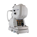
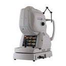
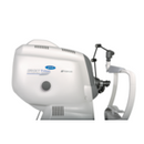
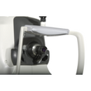
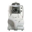
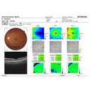
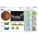
The Topcon DRI OCT Triton is a multi-modal swept source OCT with a non-mydriatic color fundus camera. Utilizing a 1,050 nm wavelength light source, and a scanning speed of 100,000 A Scans/sec, it provides uniform scanning sensitivity allowing superior visualization of the vitreous and choroid in the same scan. Invisible OCT scanning light, eye tracking during capture of selected scans, along with high scanning speeds reduce the effect of patient eye movement, improving workflow and allowing for more data to be collected in a shorter period of time. A 12 mm x 9 mm widefield scan along with automated layer segmentation provides measurement and topographical maps, with reference database, of the optic nerve and macula in one scan.
The Triton Plus model also includes a monochrome camera for fluorescein angiography and fundus auto fluorescence utilizing the exclusive Spaide auto fluorescence filters.
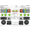
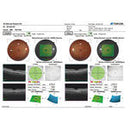
The easy-to-use, intuitive IMAGEnet 6 software enables dynamic viewing of the OCT data, providing 3D, 2D and fundus images simultaneously. Pin-Point™ Registration identifies an exact pathological location across all imaging modalities available within the Triton. In addition, both compare and trend analysis functions allow users to view serial exams of several scan protocols including our 12x9mm 3D Wide field scan. En face technology*, with layer flattening application allows for visualization of the various layers of the retina.
*Requires IMAGEnet 6 software.
Key Features:
- 100,000 A Scan/sec Swept Source OCT
- Significantly reduces capture time compared to conventional OCTs
- Reduces the impact of eye movement
- Allows for more OCT data to be collected
- Multimodal Swept Source OCT with 1050 nm light source for posterior and anterior segment OCT, color, and red free imaging. FA and FAF photography available on the Plus model.
- Saves space
- Improves workflow by reducing testing time
- Reduces costs and improves ROI
- Swept Source OCT with 1 micron wavelength light source
- Allows deeper penetration into choroid and sclera
- Less light scattering, improves results in eyes with cataracts and other media opacities
- Provides uniform sensitivity at both the top and bottom of the scanning window, allowing superior visualization of vitreous and choroid in the same scan
- Allows for scanning of highly myopic patients and patients whose pathology cannot be captured with conventional SD OCT
- Invisible OCT scanning light and high imaging speed of 100,000 A Scans/sec reduce effect of eye movements and allows more data to be collected per scan
- Widefield OCT, 12 mm x 9 mm scan captures the macula and disc in the same scan. Includes reference database of Total Retina, RFNL, GCL + IPL, GCL + IPL + RNFL thickness measurements, and optic disc parameters
- Automatic layer segmentation utilizing the Topcon Advanced Boundary Software (TABS™)
- High Density 512 x 256 OCT scanning pattern captures twice as much OCT data as conventional 512 x 128 scanning patterns, significantly increasing the available data for diagnosis
- Extensive Reference Database of RNFL, Total Retinal, GCL + IPL, GCL + IPL + RNFL thickness measurements, and optic disc parameters
- Eye Tracking comes standard with the Triton, and during capture of selected scans, ensures that you image the exact spot of the retina that you want every time.
- Pin-Point™ Registration identifies an exact pathological location across all imaging modalities available within the Triton.
- Compare and trend analysis functions allow users to view serial exams of several scan protocols including our 12x9mm 3D Wide field scan.
- IMAGEnet®6 software enables dynamic viewing of 2D, 3D and fundus images simultaneously
- Auto centering of circumpapillary grid around the disc and EDTRS grid on the fovea ensures consistent, high reproducibility results and trending data
- High resolution non-mydriatic fundus camera for color, red free and panoramic fundus imaging
- Dedicated monochrome high resolution fundus camera for fluorescein angiography and fundus auto fluorescence utilizing the exclusive Spaide auto fluorescence filters*
- En face function** with layer flattening application allows for visualization of the various layers of the retina
- *Available on DRI OCT Triton Plus model only.
- **Requires IMAGEnet 6 software.
**AVAILABLE FOR DELIVERY IN MA, NH, CT, ME, CT, RI, NY ONLY**